In this newsletter we are discussing how to recognize the signs of a calf strain along with management and treatment approaches including physical therapy.
That sudden pull in your lower leg from walking, running or jumping is quite a common problem. Depending on how badly you have injured your calf, a calf strain can either be a little nagging pain while you walk, or can take you fully out of your regular activity, and possibly require you to walk with crutches! We hope to help you learn more about the anatomy, causes, symptoms, and rehabilitation for calf strains.
Anatomy The calf is a shapely area of the body that most people know as the area that works hard to lift you up onto your toes and help you jump. Few people are aware, however, that there is more than one muscle creating the bulge of the calf area.
The large muscle that makes up the bulk of the calf muscle, and the one that most people are aware of, is called the gastrocnemius muscle. This muscle spans from the back of your heel on one end (the Achilles tendon) to the lower end of your thighbone on the other end. Since this muscle crosses both at the ankle joint and at the knee joint, it has functions for both these joints. The gastrocnemius muscle points the ankle downward, or in medical terms, plantar-flexes the ankle. This action is used to raise you up onto your tippy toes and to push yourself up as you jump.
The gastrocnemius’ function around the knee is to help bend the knee, which assists to unlock the knee from a perfectly straight knee position. There are two distinct portions of the gastrocnemius muscle; which are termed the medial and lateral heads. In a well-defined calf, these two heads can easily be seen from behind as the subject rises up onto their toes. The medial head extends farther down the calf than the lateral head.
The second muscle making up the calf is the soleus muscle. This muscle is a long, flat muscle that lies behind the gastrocnemius muscle, and directly along the back of the shin (tibia) bone. The soleus muscle has a common attachment with the gastrocnemius muscle to the back of the heel (Achilles tendon.) It does not cross over the knee joint but rather attaches simply to the back of the shinbone and the back of the bone on the outside of the lower leg, called the fibula. Together the two heads of the gastrocnemius muscle along with the soleus muscle are collectively referred to as the triceps surae.
The soleus muscle functions in conjunction with the gastrocnemius muscle to plantar flex your foot. When the knee is bent, the gastrocnemius is biomechanically at a disadvantage so it is the soleus muscle that is the main muscle to perform the plantar-flexion action. For instance, when you sit in a car and press the gas or brake pedals it is the soleus muscle hardest at work. In addition to plantar-flexing the foot, one of the other most important functions of the soleus muscle is to help us with our daily upright posture by resisting us from swaying too much in the forward direction; if the soleus muscle wasn’t constantly at work, we would fall over!
Together the muscles of the triceps surae work particularly hard when walking. When the heel strikes the ground these muscles work to decelerate the lower leg and resist it from moving too far forward. As the step progresses, these muscles work to plantar-flex the foot and get it ready to propel forward for the next step.
The final muscle in the back of the calf is called the plantaris muscle. It is a very small, thin, rope-like muscle, which is located more to the outside of the calf area. This muscle crosses over both the knee and ankle and also attaches into the Achilles tendon. This muscle works in conjunction with the gastrocnemius and the soleus to plantar-flex the foot.
What causes a calf strain?
A calf strain, or a pulled calf occurs when a muscle in your calf is overstretched or overworked. Even if the injury from overstretching or overworking occurs more to the attaching tendon at the top or bottom of the calf muscles it is still classified under the term calf strain. A calf strain can occur due to a one-time overstretching or overworking of the calf (acute injury) or it can occur from repetitive use of the calf over time (overuse injury).
Although strains can occur in any of the three calf muscles, they most often occur in the large gastrocnemius muscle due to its size and the fact that it crosses two joints (the knee and the ankle). More specifically, it is the medial head of the gastrocnemius that most often sustains a muscle strain. The next most common strain is to the soleus, and then to the plantaris muscle. A calf strain often occurs when the calf muscles are working eccentrically (working while under a stretch), such as coming down from a jump, and also during the time when you are about to push off to jump again. Most often the strain occurs at the musculotendinous junction of one of the muscles (where the muscle attaches to its tendon) but it can occur anywhere along the muscle belly as well.
As we age, the tissues of the body lose some of their elasticity, including the muscles and tendons. For this reason, strains, including calf strains, are more common in the active middle-aged patient. In addition to the tissue changes, often these people are ‘weekend warriors’ who do little to keep their muscles flexible and strong throughout the week but aggressively do sport on the weekend, which also puts them at greater risk.
How are calf strains classified?
There are several classification systems developed and in use regarding general muscle strains but the most commonly used system includes three grades. These grades can be used when describing a calf strain. It should be noted that all muscle strains include tearing of some muscle fibers:
Grade I (mild): Very few muscle fibers have been injured. Pain may not be felt until the day after the instigating activity. Strength and range of motion of the calf remains full but can be sore, and no swelling or bruising is noted.
Grade II (moderate): This is the largest and most variable category. In this category many muscle fibers are torn, which results in a decrease in plantar-flexion strength and often a limited range of motion going into the other direction (pulling the foot upwards, or dorsi-flexion). Some muscle fibers remain uninjured and intact. Pain is present both when stretching the calf and on muscle strength testing. Swelling and bruising are common.
Grade III (severe): All fibers of the affected calf muscle are completely torn. This means that the muscle is completely torn into two parts or the muscle belly has torn from its attachment to the tendon. Severe swelling, pain, and bruising accompany a grade III strain. It’s hard to generate any force on strength testing of plantar-flexion due to the tear, however the other uninjured calf muscles may compensate to initiate some strength. Range of motion is severely limited due to pain.
What does a calf strain feel like?
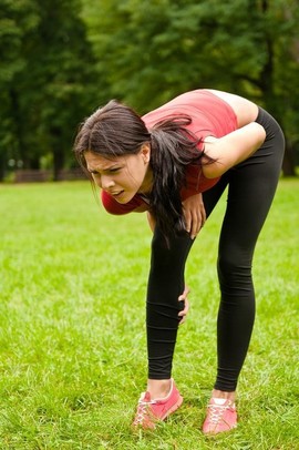
Several symptoms can indicate that you have incurred a calf strain. The symptoms you feel will depend on the grade of strain you have incurred:
● sudden onset of pain, or pain/soreness that comes on the next day related to a specific event
● muscle spasm in the area
● stiffness or tightness in the area
● symptoms aggravated by standing on your toes, walking or jogging
● pain on touching the injured calf area
● mild, moderate, or severely limited range of movement when trying to pull your foot up or stretch your calf
● decreased strength in the injured muscle
● bruising or discoloration in the area or in your ankle or foot (gravity carries the bruising down your limb)
● local swelling in the area or in your ankle or foot
● a "knotted up" feeling
● a feeling of being kicked or hit in the back of the leg (usually a severe strain or may also occur with an Achilles tendon rupture)
● hearing a “pop” when the injury occurs (usually also a severe strain or again may occur with an Achilles tendon rupture)
● a local divot or bump in the affected area due to the torn muscle fibers (usually with a severe grade II or a grade III strain)
Rehabilitation
The initial approach to physical therapy of your calf strain will depend on how long after your injury that you seek treatment. The immediate line of defense straight after sustaining a calf strain should be the application of ice and compression, followed by rest and elevation. Recent research on the benefits of applying ice immediately after an injury are beginning to be questioned as it can halt the much-needed inflammatory response, but the general consensus is still to apply ice. Applying compression as a first line of defense is extremely important. This is generally done by wrapping the affected area. Recent evidence supports the critical role of compression in preventing secondary tissue damage.
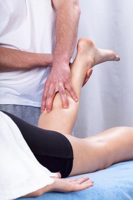
The initial aim of treatment for acute calf strains at Iron City Physical Therapy is to decrease the pain as well as any secondary inflammation in the area. Some initial inflammation is required to start the healing process, but a large inflammatory response can also lead to secondary inflammation and secondary cell injury, which affects tissues that were not directly related to the initial insult. Ice and compression can greatly assist in decreasing this detrimental secondary tissue injury. Being that the swelling from a calf injury often ends up in the ankle or foot due to gravity pulling it downwards, elevating the ankle while there is still swelling can greatly assist in moving excess tissue fluid back towards your heart and out of your limb.  Inflammation that remains after the initial few days of healing is undesired, so by this stage of healing, the aim is to finally eliminate any remaining swelling. Massage of the injured area or the tissues surrounding the area may be helpful in both decreasing swelling and decreasing pain. Depending on the severity of the strain and the time that has elapsed since the injury, massage directly over the torn calf muscle can slow the healing process and may lead to other muscle complications so be sure to heed your physical therapist’s advice on whether or not this is something you should be doing on your own.
Medication to ease the pain or inflammation can often be very beneficial in the overall treatment of a calf strain.
Depending on the degree of your strain and stage of healing, your physical therapist may suggest you see your doctor to discuss the use of anti-inflammatory or pain-relieving medications in conjunction with your physical therapy treatment. Your physical therapist may even liaise directly with your doctor to obtain their advice on the use of medication in your individual case.
Once the initial pain and inflammation has calmed down, your physical therapist will focus on improving the range of motion and strength of your calf. Early static stretches to increase the movement of your calf muscle encourage the healing tissues to withstand being loaded, and they ensure that you do not lose any range of motion. As your range of motion improves, more aggressive stretches will be added, however stretching should be limited such that it never causes pain.
Feeling a gentle stretch at the end of the range of motion should be the limit otherwise further damage could occur to the calf muscle. As the muscle advances in the healing process, dynamic stretching (rapid motions that stretch the tissues quickly) will also be taught and will be incorporated into your rehabilitation exercise routine in order to prepare your calf to return to more taxing movements such as prolonged walking, stair climbing, and jumping.
Dynamic stretches are used to prepare the tissues for activity whereas static stretches focus more on gaining flexibility.
Rest is also an important part of your physical therapy treatment. ‘Relative rest’ is a term used to describe a scale of resting compared to the normal activity you would be doing. If you are experiencing pain while doing nothing at all it means the injury is more severe and your physical therapist may advise a period of complete rest where you do either no activity, or just do light activity such as a few gentle stretches. As your pain improves then the rest to activity balance will swing the other way such that you will still require more rest for the calf than usual, but there will also be a gradual increase in activity including more aggressive stretches along with strengthening as long as there is no return in symptoms.
A long with stretching exercises, your physical therapist will also prescribe strengthening exercises in order to get your calf back in top shape. Initially your therapist may suggest that you only do isometric contractions of your muscle, which means that you tighten the affected muscle without actually moving the associated joints. An example of this type of contraction may be sitting with your foot flat against a wall, and pushing into the wall without actually moving your ankle. This type of contraction is an effective way to begin strengthening your calf. As the muscle continues to heal, more aggressive strengthening will be prescribed where you are raising up onto your toes with part or all of your body weight. When appropriate your therapist will prescribe strengthening exercises with free weights, elastic bands or tubing, weight machines, or cardiovascular machines such as stationary bicycles or a treadmill in order to continue to increase the strength and endurance in your calf. As your calf is more fully healed, your therapist will add eccentric type strengthening to your rehabilitation program. Eccentric exercises are ones that put load through your muscle as it is lengthening. These types of exercises are necessary as part of your rehabilitation program in order to prepare your calf for the return to normal everyday activity and sport. Lowering yourself off the end of a step or landing from jumping are examples of eccentric exercises for your calf. Quite often it is an eccentric contraction of the muscle that has caused the calf strain in the first place, so training the muscle (when the time is right) to withstand this type of force is crucial to ensuring it won’t be re-injured. Plyometrics is a form of power strengthening that is a particularly important part of the end stage of your rehabilitation for a calf strain, especially if you are involved in sport. Plyometrics involves repetitive jumping which forces your calf muscle to engage in force as it repetitively shortens and lengthens. This type of training maximally loads the calf muscle.
In addition to stretching and strengthening the muscle, taping, wrapping or using a soft support/brace on the calf may be suggested by your physical therapist in order to assist initial swelling, and to provide support to the muscle as you rehabilitate it. They may even teach you how to tape or wrap your own muscle so you can do it on your own.
A critical part of our treatment for a calf strain at Iron City Physical Therapy includes advice on finally returning to your full normal physical activity level. A calf strain can easily be aggravated if too much stress is put through it at an inappropriate time. Calf strains can easily be re-aggravated. Returning to your normal physical activity at a graduated pace is key in avoiding a repetitive calf strain. Advice from your physical therapist on the acceptable level of activity at each stage of your rehabilitation process will be invaluable, and will assist you in returning to your activities as quickly but as safely as possible.
In conclusion, calf strains involve a tear to the fibers of one of the calf muscles and vary in healing time depending on how severe the strain is. If you experience a calf strain, let the expert physical therapists at Iron City Physical Therapy assist you in determining the severity of your strain as well as help get you back to your everyday activity or sport by guiding you through the appropriate rehabilitation program.


